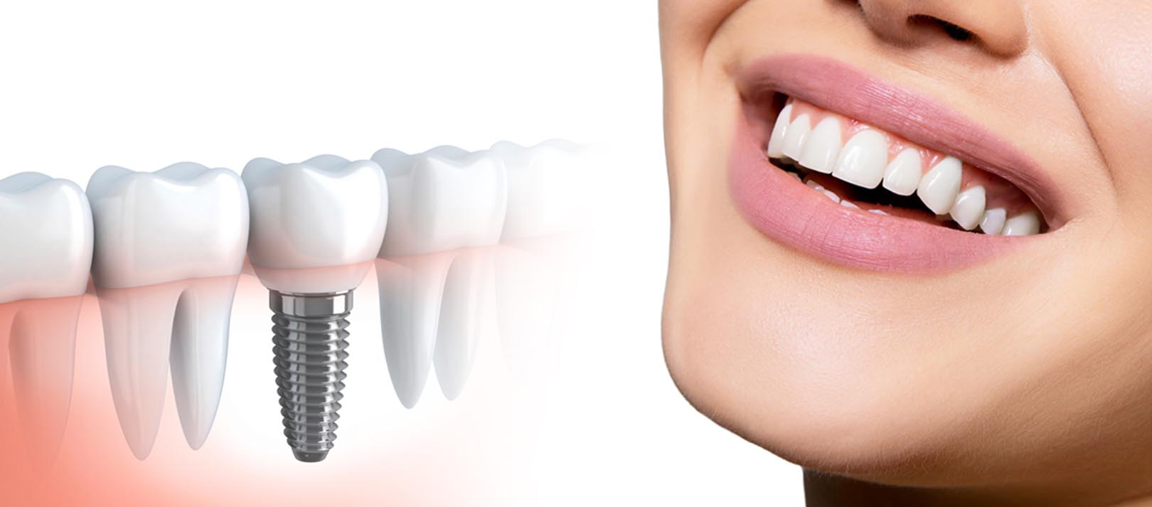
In recent years, advancements in dental technology have transformed the way dental procedures are performed, particularly in the field of dental implants. Basal implants have gained prominence among these innovations due to their ability to provide stable and reliable solutions for patients with varying bone density.
However, the success of basal implant placement relies heavily on the utilization of advanced imaging techniques. This blog will explore the crucial role of advanced imaging in basal implant placement and how it enhances treatment outcomes.
Understanding Basal Implants
Before delving into imaging techniques, it’s essential to understand basal implants. Basal implants are designed to be anchored in the basal bone—the dense, strong bone located at the bottom of the jaw—rather than the softer, cancellous bone found near the top.
This allows for immediate load-bearing capability and reduces the need for extensive bone grafting. Given the nature of this type of implant, precise planning and placement are critical for success, making advanced imaging technologies indispensable.
The Importance of Advanced Imaging
Advanced imaging technologies provide detailed insights into a patient’s dental and anatomical structure, allowing for more accurate treatment planning and execution. The primary imaging techniques used in basal implant placement include:
1. Cone Beam Computed Tomography (CBCT)
CBCT is a specialized imaging technique that provides 3D images of the jaw, teeth, and surrounding structures. This imaging method is particularly beneficial for dental implant placement due to several key advantages:
- High-Resolution Images: CBCT scans offer high-resolution images, enabling dentists to visualize bone quality and quantity in detail. This is crucial for assessing whether the basal bone can support the implant.
- 3D Visualization: Unlike traditional 2D X-rays, CBCT provides a three-dimensional view of the anatomical structures. This allows for a better spatial understanding of the jaw and helps in planning the precise positioning of the implant.
- Reduced Radiation Exposure: CBCT typically exposes patients to less radiation than traditional CT scans, making it a safer option for imaging in dental procedures.
2. Digital X-Rays
Digital X-rays are another valuable tool in the planning of basal implants. They provide quick, clear images of the dental structures, which can be enhanced and manipulated for better diagnosis. Benefits include:
- Instant Results: Digital X-rays can be viewed immediately, allowing for prompt decision-making and treatment planning.
- Enhanced Diagnostic Capability: These images can be adjusted for contrast and brightness, enabling dentists to identify potential issues that may not be visible in traditional X-rays.
3. Intraoral Scanning
Intraoral scanners create digital impressions of a patient’s mouth, allowing for the accurate modeling of teeth and surrounding structures. This technique is beneficial in several ways:
- Accuracy: Digital impressions eliminate the need for messy traditional impressions and provide highly accurate models for prosthetic design.
- Improved Patient Comfort: Intraoral scanners are less invasive and more comfortable for patients, reducing anxiety during the imaging process.
4. 3D Printing
While not an imaging technique per se, 3D printing plays a vital role in the workflow that follows advanced imaging. After obtaining detailed scans of the patient’s jaw, dentists can create precise surgical guides or custom implants using 3D printing technology.
This enhances the accuracy of the implant placement, ensuring a better fit and improved overall results.
The Impact of Advanced Imaging on Treatment Outcomes
- Enhanced Treatment Planning: Advanced imaging allows for a thorough assessment and planning before the surgical procedure. Dentists can evaluate the bone structure, identify anatomical challenges, and develop a tailored surgical plan that meets the patient’s needs.
- Increased Precision: Advanced imaging techniques provide detailed visualization, enabling dentists to place basal implants with greater accuracy. This precision minimizes the risk of complications and enhances the likelihood of successful osseointegration (the process by which the implant fuses with the bone).
- Reduced Surgical Time: Accurate pre-surgical planning and digital guides can significantly reduce the time spent in the operating room. Efficient procedures lead to better patient experiences and lower costs.
- Better Communication: Advanced imaging facilitates improved communication between dentists and patients. High-quality visuals help patients understand their condition and the planned treatment, fostering trust and confidence in the proposed procedures.
- Follow-Up and Monitoring: Advanced imaging technologies also allow for effective post-surgery monitoring of the healing process. Dentists can assess osseointegration and detect potential issues early on, ensuring timely interventions if necessary.
If you live in Kalyan, you are searching for Dentist in Kalyan. It would be best if you considered OM Dental Care. Call us to Book an Appointment: +91-9004457690.
Conclusion
The integration of advanced imaging techniques into the placement of basal implants has revolutionized the dental implant landscape. By providing detailed insights into the patient’s anatomical structure, these technologies enable precise treatment planning and enhance surgical outcomes.
As dental practices continue to embrace innovation, patients can expect improved experiences and results when opting for basal implants. If you’re considering dental implants, consult a qualified dental professional to discuss how advanced imaging can benefit your treatment journey and help you achieve the smile you’ve always desired.
